What Is a Focal Neurological Deficit | Sai Hospital, Haldwani

Uncategorized
- 14 Nov, 2025
- 254 Views
- 0 Comment
When a person experiences symptoms like sudden blackouts, seizures, frequent headaches, or unexplained confusion, neurologists often recommend an EEG test. For many patients, this term sounds unfamiliar, and it’s natural to wonder — what is EEG in neurology, and why is it important?
At Sai Hospital, Haldwani, our neurology department frequently performs EEGs to help diagnose and monitor various brain conditions. This simple, non-invasive test gives doctors a direct look into how the brain’s electrical system is functioning — something no imaging scan can fully show.
Let’s understand how EEG works, what it helps detect, and what patients can expect during the test.
What Is EEG in Neurology?
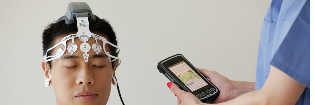
EEG stands for Electroencephalogram. It’s a neurological test that records the brain’s electrical activity using small sensors (electrodes) placed on the scalp.
Your brain constantly generates tiny electrical impulses as nerve cells communicate. An EEG captures these impulses as wave patterns, which neurologists analyze to detect any abnormal activity.
In simple terms, an EEG shows how well your brain is working electrically — much like how an ECG records heart activity.
Why Is EEG Done?
EEG helps diagnose and monitor a wide range of neurological disorders that affect brain function. Common reasons your doctor might recommend an EEG include:
1. Seizures and Epilepsy
This is the most frequent reason for EEG testing.
Abnormal bursts of brain waves during or between seizures help neurologists confirm a diagnosis of epilepsy and determine the type of seizure.
2. Loss of Consciousness or Fainting Spells
If someone experiences blackouts, confusion, or sudden unresponsiveness, an EEG helps determine if the cause is neurological.
3. Head Injuries
After trauma, an EEG can detect irregular brain wave patterns that suggest brain dysfunction or post-traumatic epilepsy.
4. Sleep Disorders
EEG helps evaluate sleep disturbances like insomnia, sleep apnea, or narcolepsy, often as part of a polysomnography (sleep study).
5. Encephalitis or Brain Infections
Abnormal EEG patterns can suggest inflammation or infection in the brain tissue.
6. Dementia and Degenerative Diseases
In conditions like Alzheimer’s disease, EEGs can reveal slowed brain activity, helping assess the disease’s progression.
7. Monitoring Brain Function in Coma
EEG is used in ICUs to track brain activity in patients with severe brain injury or coma, helping doctors assess recovery potential.
How Does EEG Work?
At Sai Hospital, Haldwani, the EEG procedure is performed by trained technicians under neurologist supervision. Here’s what typically happens:
- Preparation:
The patient is comfortably seated or lying down. The scalp is cleaned, and small electrodes are attached using a conductive paste or cap. - Recording Brain Waves:
The electrodes detect the brain’s natural electrical signals. These signals are amplified and displayed as wave patterns on a computer screen. - Testing Conditions:
Patients may be asked to:- Open and close their eyes
- Take deep breaths (hyperventilation test)
- Look at flashing lights (to test seizure sensitivity)
- Relax or even sleep during the recording
- Duration:
A standard EEG takes 30 to 45 minutes, though longer sessions or sleep EEGs may last up to several hours. - After the Test:
Once the recording is done, the electrodes are removed, and the neurologist analyzes the patterns for abnormalities.
The test is completely painless, safe, and non-invasive — there are no shocks or risks of side effects.
Understanding EEG Results
An EEG report shows brain activity as waveforms — alpha, beta, delta, and theta waves — each representing different mental states like relaxation, alertness, or sleep.
Normal EEG:
Displays consistent, symmetrical wave patterns appropriate for the patient’s age and state (awake or asleep).
Abnormal EEG:
May show:
- Spike or sharp waves – common in epilepsy
- Slowed activity – seen in encephalitis, brain injury, or metabolic disorders
- Lack of activity – may indicate deep coma or brain death in severe cases
Neurologists at Sai Hospital carefully interpret these findings alongside clinical symptoms and other test results before making a diagnosis.
Types of EEG Tests
Depending on your condition, your neurologist may recommend one of the following:
1. Routine EEG
Standard 30–60-minute recording done in an outpatient setting.
2. Sleep-Deprived EEG
The patient is kept awake for several hours before the test to increase the chances of detecting abnormal brain activity.
3. Ambulatory EEG
A portable device records brain activity for 24–72 hours while the patient goes about daily activities.
4. Video EEG Monitoring
Used for epilepsy diagnosis and pre-surgical evaluation — continuous EEG is recorded along with video to capture seizure events and behavior.
5. Intraoperative EEG
Performed during neurosurgery to monitor brain function and prevent damage to critical areas.
What to Expect Before and After the Test
Before the Test
- Wash your hair to remove oil or hair products.
- Avoid caffeine or stimulants 8–12 hours before the test.
- Continue medications unless your doctor advises otherwise.
After the Test
- You can resume normal activities immediately.
- The neurologist reviews your EEG results and discusses findings during follow-up.
If the EEG indicates abnormal brain activity, further imaging tests like MRI or CT scan may be advised for detailed evaluation.
Benefits of EEG
- Early detection: Identifies epilepsy, brain inflammation, or functional changes before structural damage appears on scans.
- Safe and non-invasive: No radiation or discomfort.
- Real-time insight: Shows active brain function, unlike MRI which shows structure.
- Guides treatment: Helps neurologists track progress or medication response in seizure disorders.
Limitations of EEG
While EEG is highly valuable, it has some limitations. It cannot always pinpoint the exact cause of abnormalities or detect deep-brain lesions. Therefore, neurologists often combine EEG with other tests like MRI, CT, or NCV for comprehensive analysis.
EEG at Sai Hospital, Haldwani
Sai Hospital’s Department of Neurology offers state-of-the-art EEG facilities managed by experienced neurologists and technicians.
We ensure:
- Accurate recordings using modern digital EEG systems
- Comfortable, patient-friendly environment
- Expert interpretation and reporting within a short turnaround time
- Combined evaluation with MRI or NCV when needed
Whether it’s for diagnosing seizures, sleep disorders, or monitoring recovery, Sai Hospital provides complete neurodiagnostic support under one roof.
FAQs: What Is EEG in Neurology
1. Is EEG painful?
No, EEG is a painless, non-invasive test that simply records electrical activity through scalp sensors.
2. How long does an EEG take?
A standard EEG takes about 30–45 minutes, while extended tests can last several hours.
3. Can EEG detect all brain problems?
EEG detects functional abnormalities like seizures or brain wave disruptions but may not identify structural issues — for that, imaging tests are used.
4. Do I need to stop medications before an EEG?
Only if advised by your doctor. Some drugs can affect results, so always inform your neurologist about current prescriptions.
5. Is EEG safe for children and elderly patients?
Yes, EEG is completely safe and suitable for all age groups, including infants and older adults.
Conclusion
So, what is EEG in neurology? It’s a vital diagnostic tool that records the brain’s electrical activity to help detect and manage neurological conditions like epilepsy, sleep disorders, or brain injury.
At Sai Hospital, Haldwani, we use advanced EEG technology to ensure accurate diagnosis, early detection, and better treatment planning for every patient.
If you’re experiencing unexplained blackouts, seizures, or neurological symptoms, consult our neurology experts for a thorough evaluation — because understanding your brain’s signals is the first step to protecting your health.